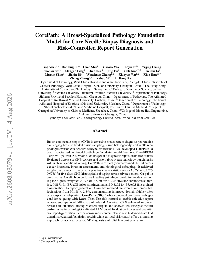
Breast core needle biopsy (CNB) is central to breast cancer diagnosis yet remains challenging because limited tissue sampling, lesion heterogeneity, and subtle morphologic overlap can obscure subtype distinctions. We developed CorePath, a breast-specialized multimodal pathology foundation model fine-tuned from PRISM using 7901 paired CNB whole-slide images and diagnostic reports from two centers. Evaluated across six CNB cohorts and two public breast pathology benchmarks without task-specific retraining, CorePath consistently outperformed PRISM across cancer detection, invasion assessment, and histological subtyping. It achieved weighted area under the receiver operating characteristic curves (AUCs) of 0.9526-0.9735 for five-class CNB histological subtyping across private centers. On public benchmarks, CorePath outperformed leading pathology foundation models, achieving the highest weighted AUCs of 0.7780 for BCNB invasive carcinoma subtyping, 0.8178 for BRACS lesion stratification, and 0.8252 for BRACS fine-grained classification. In report generation, CorePath reduced the overall non-breast hallucinations from 30.1% to 2.8%, demonstrating improved domain fidelity after breast-specific adaptation. CorePath-CRG further combined conformal subtype-confidence gating with Learn-Then-Test risk control to enable selective report release, subtype-level fallback, and deferral. CorePath-CRG achieved zero non-breast hallucinations among released outputs and showed the strongest overall performance in pathologist-validated LLM-based Evaluation Scores and quantitative report-generation metrics across most centers. These results demonstrate that domain-specialized foundation models with statistical risk control offer a promising approach for accurate breast CNB diagnosis and reliable report generation.
CorePath: A Breast-Specialized Pathology Foundation Model for Core Needle Biopsy Diagnosis and Risk-Controlled Report Generation
Breast core needle biopsy (CNB) is central to breast cancer diagnosis yet remains challenging because limited tissue sampling, lesion heterogeneity, and subtle morphologic overlap can obscure subtype distinctions.
- Preview
- Year
- 2026
- Hosting
- Excerpt onlyCC-BY-NC-4.0
Cite
Notes
Only stored in your browser.
Attribution
- Abstract & full text
- arxiv.org/abs/2608.03079CC-BY-NC-4.0
- TL;DR
- Semantic Scholar